Biceps tendinopathy (Canine)
What is Biceps tendinopathy (Canine)?
The shoulder joint of the dog is composed of the glenoid cavity (on the scapula or shoulder blade) and the humeral head (the top of the upper arm bone). There are many soft tissue structures in this region that confer stability and cover to the shoulder. Paired glenohumeral ligaments provide static stability, with multiple surrounding muscles supplying dynamic stability. These muscles include the supraspinatus, infraspinatus, teres minor, subscapularis, and biceps brachii. The tendon of the biceps passes over the front of the shoulder joint and is susceptible to injury through overuse and trauma (direct or indirect), leading to inflammation of the tendon and its surrounding sheath.
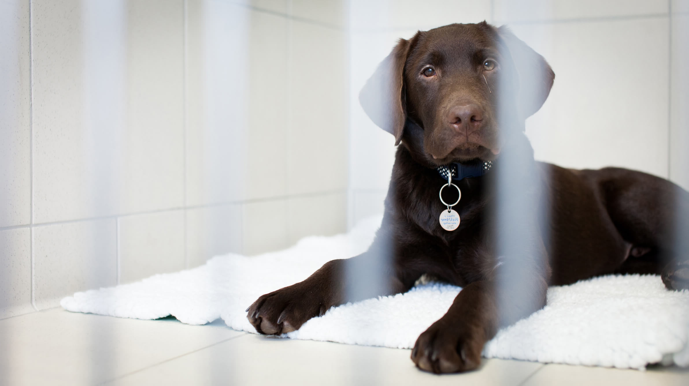
Biceps tendinopathy (tendon injury) is a condition seen in dogs, affecting middle-aged to older, medium to large-sized breeds. Labrador retrievers and Rottweilers tend to be over-represented with this condition.
What are the signs of biceps tendinopathy?
The clinical signs of biceps tendinopathy are a slow insidious lameness of one or both (can be bilateral) forelimbs. Affected dogs tend to present with chronic forelimb lameness that may shift from one forelimb to the other (in bilateral cases), and is usually worse following exercise. Some dogs may be completely non-weightbearing if the tendinopathy is severe.
How is biceps tendinopathy diagnosed?
On clinical examination pain on manipulation and palpation can be seen. If the biceps tendon has completely failed there will be excessive flexion in the shoulder. Investigation via shoulder radiography and ultrasound can provide a definitive diagnosis in some cases. Others require more advanced imaging.
Radiography is useful to exclude any concurrent shoulder injury and can show obvious mineralisation within the bicipital groove (where the biceps tendon inserts). Ultrasound allows assessment of the soft tissue components of the biceps tendon and comparison of pathology from one shoulder to the other. Disruption of the normal tendon structure can be seen by experienced ultrasonographers.
In the more challenging cases, advanced imaging via CT/MRI may be required. Arthroscopy (keyhole surgery for direct assessment) is often necessary for confirmation.
How is biceps tendinopathy treated?
Treatment for biceps tendinopathy is initially conservative, through rest and medical management. A period of rest along with anti-inflammatory medication is trialled. This is to allow time for the tendon to heal and inflammation to settle. Return to exercise too early can lead to chronic lameness or complete tendon rupture.
Some cases do not respond to conservative management and require surgery for tendon transection (done via arthroscopy). Other treatment modalities have been trialled such as extracorporeal shockwave therapy with potentially promising results.
What is the prognosis for biceps tendinopathy?
The outcome for this condition is dependent upon several factors – the severity and chronicity of the tendinopathy, rest and restriction during management or the postoperative period, weight control and the degree of associated degenerative joint disease already present. Most cases go on to achieve excellent outcomes, with the majority of dogs returning to limb function within 3 months; others may take longer (9 months or so in some patients).
Stay in touch
Follow us on social media and keep up to date with all the latest news from the Grove clinic.